


HTD™ Nucleolus Stains
Description
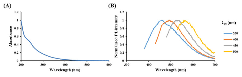
The amphiphilic HTD™ nucleolus targets stains label nucleolus efficiently. The stains are used for labeling the nucleolus of live adherent and suspension cells, as tracers for the dynamics of nucleolus and for multi-generational cellular tracing. HTD™ nucleolus stains have full-color fluorescence characteristics, which emit blue, green, and red fluorescence under UV, blue, and green light excitations, respectively.
Features
- Full-color fluorescence, HTD™ nucleolus stains have full-color fluorescence characteristics, which emit blue, green, and red fluorescence under UV, blue, and green light excitations, respectively.
- Application, HTD™ nucleolus stains are applied to nucleolus of live, dead, or fixed cells.
- Multi-generational tracking, the stains are used for labeling the nucleolus structures of live adherent and suspension cells, as tracers for the dynamics of nucleolus and for multi-generational cellular tracing.
- Efficiency, incubate the HTD™ Nucleolus stains for 1 minute, then proceed to image cells.
- Photostability. more than 9 hours in live cells; more than 15 minutes in specimen.
- Low cytotoxicity, does not affect viability or proliferation.
Performance
- Multi-generational tracking
![]()
HTD™ nucleolus enabled long-term nucleolus tracking in cells. The stain was transferred to daughter cells by dilution of the parent cell, and the staining pattern of HTD™ nucleolus remained in nucleolus for at least six days after staining.
- Efficiency (Staining time)

- Staining in fixed and permeabilized cells

- Photostability – Co-stained with Hoechst

HTD™ nucleolus and Hoechst nucleus were co-stained and irradiated under UV light (λex: 360-370 nm) for 12 minutes. Observed that Hoechst nucleus completely photobleaching only HTD™ nucleolus still photostability.
- Low cytotoxicity

Spectural Properties
- Blue fluorescence channel:Ex/Em: 360-370 / ≥ 420 nm
- Green fluorescence channel:Ex/Em: 460-490 / ≥ 520 nm
- Red fluorescence channel:Ex/Em: 525-545 / ≥ 575 nm
Protocol Overview
